🎯
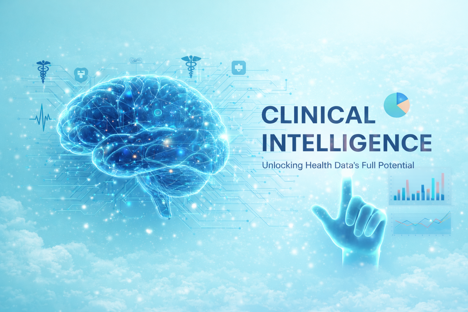
Engine 1 of 4 • Annotation Platform

OmniLabelX

Intelligent Medical Image Annotation

Transform your medical imaging workflow with AI-powered annotation. OmniLabelX combines advanced pre-labeling algorithms with intuitive tools to reduce annotation time by 70% while improving consistency and accuracy across your team.

70%
Time Reduction
80+
Features
15+
Modalities
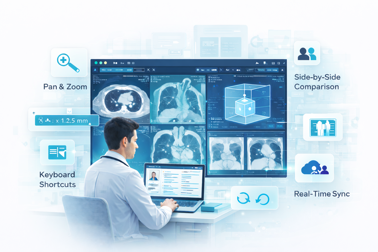
OmniLabelX — Annotation Canvas
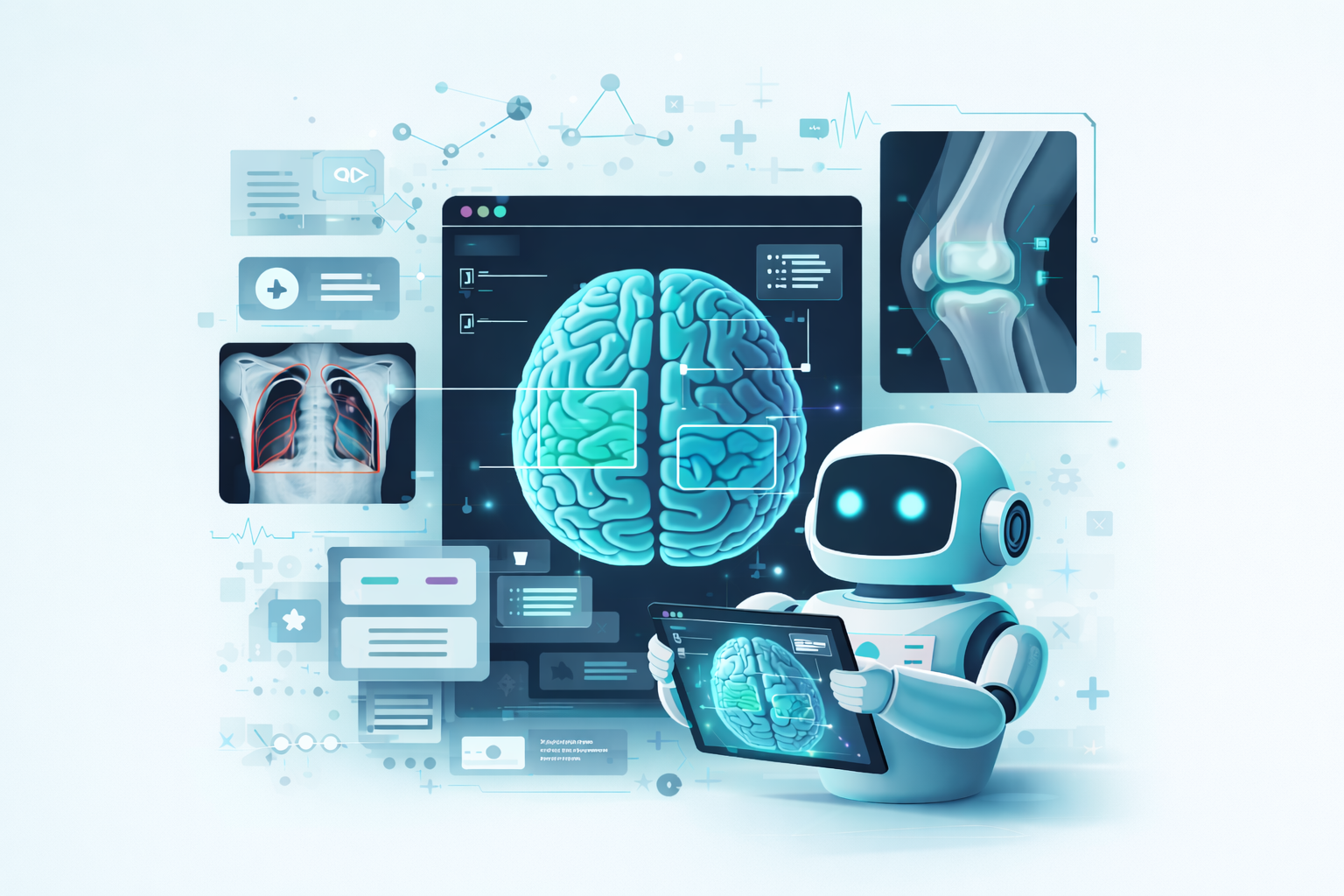
OmniLabelX Annotation Canvas
AI Pre-labeling
SAM + YOLOv8
📊
Accuracy
0.88 Dice Score

Medical Image Annotation is Time-Consuming and Inconsistent

Radiologists and clinicians spend countless hours manually annotating medical images — drawing contours, marking lesions, and documenting findings. This repetitive work leads to fatigue, inconsistency, and takes valuable time away from patient care.

Traditional annotation tools weren't built for the complexity of medical imaging. They lack DICOM support, can't handle 3D volumes, and offer no AI assistance to accelerate the workflow.

  • Average 40 minutes per complex study for manual annotation
  • Inter-annotator variability of 15-20% on challenging cases
  • No standardized export formats for AI training pipelines
  • Limited support for multi-modal and 3D imaging
OmniLabelX Annotation Canvas

Everything You Need to Annotate Medical Images

From simple bounding boxes to complex 3D volumetric segmentation, OmniLabelX provides a comprehensive toolkit built specifically for healthcare imaging.

✏️

Comprehensive 2D Tools

Full suite of annotation tools including bounding boxes, polygons, polylines, freehand drawing, brush/eraser, and smart scissors. Keyboard shortcuts for rapid annotation.

🧊

3D Volumetric Segmentation

MONAI-powered 3D segmentation for CT and MRI volumes. Annotate slice-by-slice with automatic propagation, or use AI-assisted whole-volume segmentation.

🤖

AI-Powered Pre-labeling

Integrated SAM (Segment Anything Model) and YOLOv8 generate initial annotations automatically. Click once to segment — AI does the rest. Reduce manual work by 70%.

👥

Multi-User Collaboration

Real-time collaboration with role-based access control. Annotators, reviewers, and approvers work in a structured workflow with full audit trail.

📋

Quality Assurance

Built-in QA with inter-annotator agreement metrics, Dice coefficient tracking, and gold standard validation. Ensure consistency across your annotation team.

📤

Standards-Compliant Export

Export to COCO JSON, NIfTI, DICOM-SR, Pascal VOC, and FHIR Observation. Ready for AI training pipelines or clinical documentation.

📐

Measurement Tools

Precise measurement capabilities including length, area, volume, angle, and Hounsfield unit analysis. RECIST-compliant tumor measurements.

🏷️

Structured Labeling

SNOMED-CT and RadLex ontology integration. Create custom label hierarchies with attributes, relationships, and clinical context.

🔒

De-identification

Automatic PHI removal from DICOM headers using RSNA profiles. Neural network-based burned-in text detection and redaction.

How OmniLabelX Works

A streamlined four-step workflow that takes you from raw DICOM to AI-ready annotations.

1

Upload

Import DICOM from PACS via DICOMweb or upload directly. Automatic metadata extraction, validation, and optional de-identification.

2

Pre-label

AI models analyze the image and generate initial annotations. SAM segments structures, YOLOv8 detects findings. Review and accept or refine.

3

Refine

Use the annotation canvas to perfect labels. Add measurements, apply ontology codes, and collaborate with team members in real-time.

4

Export

QA validation ensures quality. Approval workflow captures sign-off. Export in your preferred format — COCO, NIfTI, DICOM-SR, or FHIR.

Intuitive Interface, Powerful Capabilities

Built on Cornerstone.js and Fabric.js, OmniLabelX provides a responsive, feature-rich annotation canvas that handles everything from simple X-rays to complex multi-frame CT volumes with thousands of slices.

  • Pan, zoom, and window/level controls with smooth performance
  • Multi-planar reconstruction (MPR) for 3D navigation
  • Side-by-side comparison of prior studies
  • Customizable hanging protocols
  • Keyboard shortcuts for every action
  • Unlimited undo/redo with full history
  • Real-time synchronization across users
See It in Action →
OmniLabelX Annotation Canvas

Supported Imaging Modalities

OmniLabelX works with all major medical imaging formats and modalities used in clinical practice.

🩻
CT Scan
Computed Tomography
🧠
MRI
Magnetic Resonance
📷
X-Ray
Radiography
📡
Ultrasound
Sonography
💫
PET/CT
Nuclear Medicine
🔬
Pathology
Whole Slide Imaging
🫀
Echo
Echocardiography
🩺
Mammography
Breast Imaging
💉
Angiography
Vascular Imaging
👁️
Fundus/OCT
Ophthalmology
🔭
Endoscopy
GI Imaging
🦴
DEXA
Bone Densitometry

Real-World Use Cases

See how healthcare organizations use OmniLabelX to accelerate their imaging workflows.

🫁

Lung Nodule Detection Training

A diagnostic chain annotated 50,000 chest CT scans to train an AI model for lung nodule detection. OmniLabelX's AI pre-labeling reduced annotation time from 45 minutes to 12 minutes per study.

73% time saved
0.91 Dice score
🧠

Brain Tumor Segmentation

A neuro-oncology research team used 3D volumetric tools to segment gliomas across 2,000 MRI studies. Multi-user collaboration enabled parallel annotation with consistent quality.

throughput
8% inter-annotator variance
🔬

Digital Pathology QC

A pathology lab deployed OmniLabelX for whole slide image annotation. Gigapixel image support and AI-assisted cell detection enabled rapid tumor boundary delineation.

100K× zoom support
60% faster grading
❤️

Cardiac Function Analysis

A cardiology department annotated echocardiograms for automated EF calculation. Frame-by-frame annotation with DICOM-SR export integrated directly with their PACS workflow.

DICOM-SR export
Real-time sync

Technical Details

Specification Details
Input Formats DICOM, NIfTI, PNG, JPEG, TIFF, SVS (pathology), OME-TIFF
Export Formats COCO JSON, Pascal VOC XML, NIfTI, DICOM-SR, FHIR Observation, CSV
AI Models SAM (Segment Anything), YOLOv8, MONAI UNet, MedSAM, custom fine-tuned models
Annotation Types Bounding box, polygon, polyline, freehand, brush, 3D mask, landmarks, measurements
Max Image Size 100,000 × 100,000 pixels (pathology), unlimited DICOM series length
Collaboration Real-time multi-user, role-based access, audit trail, version history
Ontologies SNOMED-CT, RadLex, ICD-10, custom hierarchies
Standards DICOM, DICOMweb (WADO-RS, STOW-RS), FHIR R5, NDHM/ABDM
De-identification RSNA CTP profiles, neural network burned-in text detection, face detection
Browser Support Chrome 90+, Firefox 88+, Safari 14+, Edge 90+

Works Seamlessly with Other Engines

OmniLabelX annotations flow directly into OmniReasonX for report generation, OmniModelX for predictive analytics, and OmniWeaveX for federated learning.

🧠
OmniReasonX
Generate reports from annotations
📊
OmniModelX
Train predictive models
🔗
OmniWeaveX
Federated model training

Ready to Accelerate Your Annotation Workflow?

See how OmniLabelX can reduce your annotation time by 70% while improving quality and consistency. Schedule a personalized demo with your own imaging data.